Bioimaging
Bioimaging
It is our mission to facilitate science and provide access to know-how and measurement time on state-of-the-art microscopes. We have ample devices available, including numerous confocal microscopes and super-resolution systems, slide scanners, high content screening, and 3D whole organ imaging (e.g. light sheet microscopes) as well as transmission and scanning electron microscopes, light and electron microscopy-combined systems and block face scanning electron microscopy solutions. We take pride in our support for image analysis both with infrastructure and expertise.
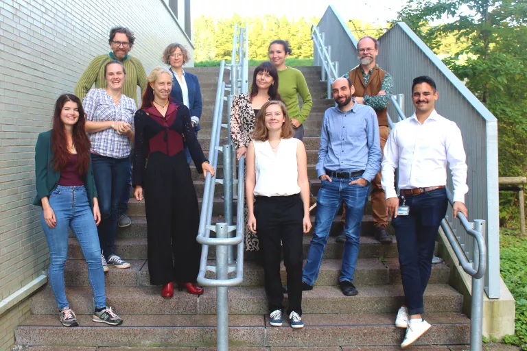
The VIB Bioimaging Core Leuven is headed by Pablo Hernández Varas (pablo.hernandezvaras@vib.be).
VIB Bioimaging Core
The VIB Bioimaging Core in Leuven supports imaging from in vitro to in vivo, from the meso to the Ångström scale, and from morphology to mechanism. They have ample solutions available, including confocal microscopes, slide scanning, high content screening, and 3D whole organ imaging.
Let's show you around!
Get to know some of the incredible tools and people driving the Bioimaging Core through this vlog with Pablo Hernández Varas, head of the VIB Bioimaging Core Leuven. (Find out more about Pablo, his career path and what lead him to joining VIB in the blog post below!)
Take a Virtual Tour
Want to see our microscopes? Check out what we have available at the VIB-KU Leuven Center for Neuroscience via our 360° virtual tour.
In the Spotlight
Check out some recent collaborations between the VIB Bioimaging Core Leuven and labs at the VIB-KU Leuven Center for Neuroscience.
TDP-43 seeding induces cytoplasmic aggregation heterogeneity and nuclear loss of function of TDP-43
Neuron (2025) in collaboration with the Sandrine Da Cruz Lab
A postsynaptic GPR158-PLCXD2 complex controls spine apparatus abundance and dendritic spine maturation
Dev Cell (2025) in collaboration with the Joris De Wit Lab
Soma-localized Rab39 inhibits synaptic autophagy by controlling trafficking of Atg9 vesicles
EMBO J (2025) in collaboration with the Patrik Verstreken Lab

We are a Nikon Center of Excellence
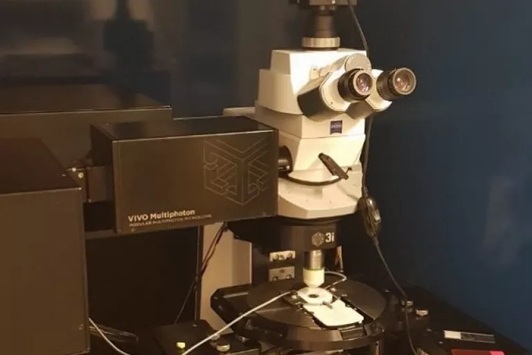
In Vivo Advanced Imaging